Atopic Dermatitis or Subacute Cutaneous Lupus: A Clinicopathologic Puzzle
Updated
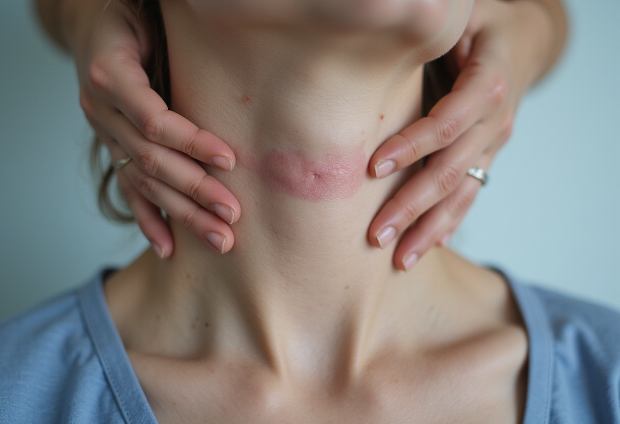
A new case report published in Cureus documents how a presentation that read clinically as atopic dermatitis turned out, on histologic and immunologic workup, to be subacute cutaneous lupus erythematosus (SCLE) — and it makes a pointed argument for clinicopathologic correlation whenever the two entities collide.
The diagnostic overlap is not incidental. Both diseases can present with eczematous, scaly, photosensitive patches on sun-exposed skin. Both can itch. And both can appear in patients with a history that seems, at first, to fit one label neatly. The report walks through a case in which the initial clinical impression favored atopic dermatitis, the most common chronic inflammatory dermatosis, only for subsequent evaluation to redirect the diagnosis toward SCLE, an autoimmune photosensitivity disorder in the lupus spectrum.
Why does the distinction matter so much? The therapeutic and prognostic trajectories diverge sharply. Atopic dermatitis is managed with topical anti-inflammatory agents, emollients and, in moderate-to-severe disease, biologics such as dupilumab or janus kinase inhibitors. Subacute cutaneous lupus, by contrast, is strongly associated with anti-Ro/SSA autoantibodies — present in the large majority of SCLE patients — and in a substantial minority of cases occurs in the context of systemic lupus erythematosus or drug-induced lupus, most famously linked to terbinafine, thiazides, calcium channel blockers and TNF-alpha inhibitors. Missing the diagnosis means missing the serologic workup, the medication review and the systemic evaluation that the label demands.
The case underscores several practical lessons for the clinic. First, morphology alone is an unreliable discriminator. Eczematous plaques in a photo-distributed pattern — across the shoulders, upper chest, extensor forearms, V of the neck — warrant lupus consideration even when the patient reports itch and a chronic relapsing course. Second, biopsy findings carry real weight. Direct immunofluorescence demonstrating a lupus band or particulate deposition of IgG along the dermoepidermal junction, together with interface dermatitis with necrotic keratinocytes and basement membrane thickening on hematoxylin-eosin staining, points away from the spongiosis-dominated picture of atopic dermatitis. Third, serology closes the loop: anti-Ro/SSA and anti-La/SSB antibodies, antinuclear antibody testing and, where systemic involvement is suspected, complement levels and urinalysis.
The report also flags the pharmacologic dimension. Because numerous common medications can trigger SCLE, a careful drug history — including agents started months earlier — belongs in every workup of a "treatment-resistant eczema" that behaves atypically. Persisting lesions despite appropriate atopic dermatitis therapy, distribution that follows sun-exposed sites, or histology that shows interface change rather than spongiosis should all prompt the clinician to re-examine the diagnosis rather than escalate the eczema regimen.
The authors frame their conclusion as a reminder rather than a revelation: clinicopathologic correlation remains the backbone of dermatologic diagnosis, and it is precisely in the inflammatory mimics — where one disease wears the face of another — that this discipline earns its keep. For clinicians, the takeaway is operational. Biopsy early when the clinical picture resists the expected trajectory. Pair histology with direct immunofluorescence and an autoimmune panel when photosensitive, eczematous disease refuses to respond. And treat the diagnostic label as a hypothesis under continuous test, not a settled fact.
As biologic and small-molecule options for atopic dermatitis multiply, the cost of a wrong label grows: an SCLE patient mislabeled as atopic dermatitis may receive years of ineffective escalation while the window for detecting systemic involvement narrows. Case reports like this one, by mapping the decision points in a single well-documented patient, add to the pattern library clinicians draw on when the next ambiguous presentation walks through the door.
Source: Google News: Atopic dermatitis
Amara Osei
Show full bio
Correspondent covering technology and product news in Clinical dermatology, inflammatory skin disease and biologics at Derm Field.