Vessel Patterns Under the Microscope Flag Deadly Sebaceous Skin Tumors
Updated
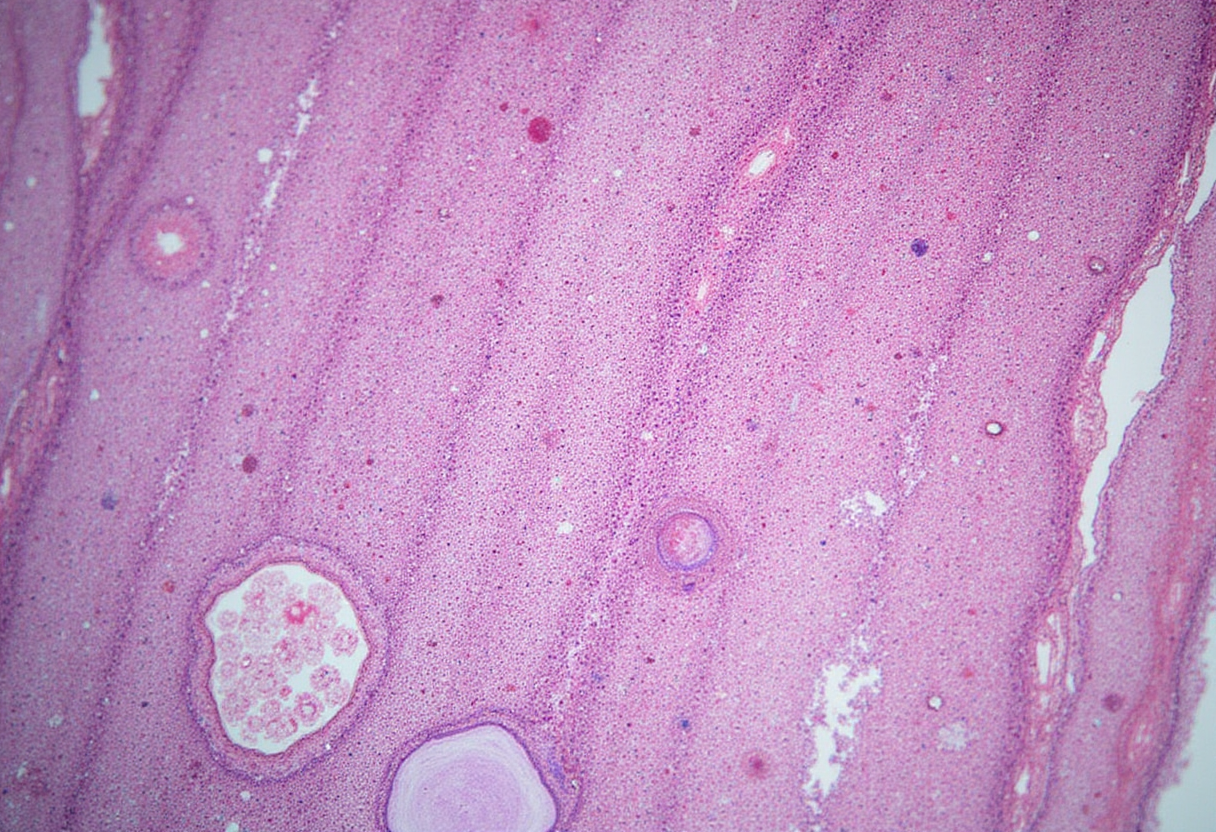
A histopathologic analysis of microvascular patterns can flag which sebaceous skin tumors turn lethal, according to work reported by Bioengineer.org. The study reframes risk assessment in sebaceous neoplasia around vessel architecture visible on routine microscopy rather than cytologic atypia alone, opening a candidate prognostic readout for tumors where grading has historically proved inconsistent.
What did the researchers actually examine?
Investigators compared the arrangement, density and morphology of intratumoral and peritumoral blood vessels across sebaceous lesions, looking for patterns that track with aggressive clinical behavior. The premise draws on a long-standing observation in solid-tumor pathology: malignant lesions recruit disorganized, leaky, often densely packed vessels, while benign counterparts tend to retain more orderly vascular networks. In sebaceous tumors, where cellular features overlap between adenomas, sebaceomas and frankly carcinomatous lesions, the vasculature may carry independent prognostic information.
Why does risk stratification matter for sebaceous tumors?
Sebaceous carcinoma remains rare but carries disproportionate mortality when it behaves aggressively, particularly in periocular sites where delayed recognition contributes to orbital invasion and metastasis. The tumor also features prominently in Muir-Torre syndrome, a Lynch-spectrum variant in which sebaceous neoplasms flag an underlying mismatch-repair defect and a heightened lifetime risk of internal malignancies. Clinicians therefore need tools that separate indolent sebaceous proliferations from lesions that warrant wide excision, sentinel-node workup, immunostaining for mismatch-repair proteins, and genetic counseling.
Current grading rests largely on architectural features such as asymmetry, depth of invasion, cytologic pleomorphism and mitotic activity. Interobserver variability is well documented, and rare sebaceous proliferations with ambiguous cytology can frustrate both dermatopathologists and Mohs surgeons planning margins.
What could change at the microscope and the bedside?
A vessel-based readout, if validated, would give dermatopathologists a second axis of assessment grounded in the tumor microenvironment. The approach would slot into existing workflows because vascular patterns can be appreciated on routine H&E sections, with optional confirmation by CD31, CD34 or ERG immunostains already in service across most dermatopathology laboratories. For clinicians, a microvascular risk score could complement staging and inform decisions about excision width, nodal surveillance and referral for mismatch-repair evaluation in patients without a known Muir-Torre diagnosis.
Key questions remain. The published source does not specify cohort size, follow-up duration, interobserver reproducibility, or whether the microvascular signature holds across periocular and extraocular sites. The threshold that separates lethal from indolent lesions, and the extent to which vessel patterns add predictive value beyond established AJCC staging, will require independent confirmation in larger, prospectively recruited cohorts.
Larger studies are now the obvious next step. If microvascular profiling proves reproducible across institutions and tumor locations, dermatopathology could gain a low-cost, immunohistochemistry-light biomarker that sharpens the line between sebaceous tumors that heal after excision and those that warrant the full oncology workup.
Source: Google News: Skin cancer
Amara Osei
Show full bio
Correspondent covering technology and product news in Clinical dermatology, inflammatory skin disease and biologics at Derm Field.