Raman Spectroscopy Plus AI Classifies BCC, SCC and Normal Skin at 84%
Updated
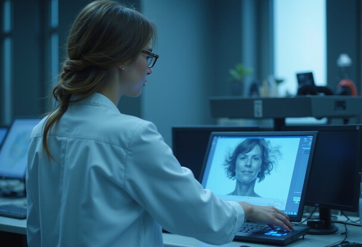
The strongest machine-learning models in a Florida Atlantic University study correctly classified normal skin, basal cell carcinoma and squamous cell carcinoma 81% to 84% of the time, using Raman spectra acquired with a handheld probe and a 785-nanometer diode laser.
The study, published in the Proceedings of SPIE as part of Advanced Chemical Microscopy for Life Science and Translational Medicine 2026, analyzed more than 50 clinical samples ex vivo — BCC, SCC and normal skin — and generated nearly 1,000 Raman spectra. Researchers then benchmarked several machine-learning classifiers against the spectral data to see how accurately the three tissue types could be separated.
The results frame both the promise and the ceiling of the approach. K-nearest neighbors and support vector machine classifiers achieved the highest overall test accuracy, at about 84%. The support vector machine reached 78.7% sensitivity and 88.6% specificity. A shallow neural network delivered 80.8% accuracy and the best receiver operating characteristic performance, with an area under the curve of 0.910. Classification of cancer versus normal tissue was stronger than discrimination between the two nonmelanoma cancer types, whose molecular signatures overlapped considerably.
That biochemical overlap has a structural explanation in the spectral data. Cancerous samples showed stronger protein-related Raman signals, while normal tissue produced stronger lipid-related signals. The spectral separation between BCC and SCC was narrower than the separation between malignant and normal tissue — the pattern that ultimately limited three-way classification accuracy.
Why dermatology needs this
Skin cancer is the most common cancer in the United States and among the most common worldwide. Nearly 1.5 million new cases were diagnosed globally in 2024, including nearly 340,000 melanomas. Nonmelanoma skin cancers — primarily BCC and SCC — are more common still, with 5.4 million cases diagnosed annually in the U.S.
Early detection drives outcomes, but many malignant lesions look similar to benign and precancerous growths on visual inspection. Biopsy followed by microscopic examination remains the diagnostic gold standard. Biopsies are invasive and costly, and clinicians sometimes excise lesions that prove benign. A rapid, non-invasive triage tool could reduce that burden.
Raman spectroscopy works differently from tissue removal. The technique measures how light scatters when it interacts with molecules in tissue, producing a molecular "fingerprint" of chemical composition without cutting or specially preparing the sample.
"The promise of this technology is that it could give clinicians another way to look beneath the surface of a skin lesion without immediately having to remove tissue," said Andrew Terentis, Ph.D., senior author and professor and chair of FAU's Department of Chemistry and Biochemistry in the Charles E. Schmidt College of Science. "By combining the molecular information provided by Raman spectroscopy with machine learning, we are beginning to see how subtle differences in tissue chemistry can be used to distinguish cancer from normal skin."
What the numbers do and don't support
An 84% three-way accuracy figure, with sensitivity below 80% for the top support vector machine model, is not yet sufficient for clinical deployment — and the authors say so directly. The work is a preliminary, ex vivo demonstration on a small sample set. It establishes that spectral patterns distinguishing normal from malignant tissue exist and are learnable by conventional classifiers, not that a handheld Raman device can replace biopsy.
"Our results are preliminary, but they point toward a future in which a rapid, non-invasive measurement could help guide clinical decisions and potentially reduce unnecessary biopsies," said Terentis.
The next steps are larger sample collections, model optimization and more advanced techniques, including deep neural networks, which the researchers expect could push diagnostic performance higher.
"Raman spectroscopy gives us a wealth of molecular information, but the challenge is teaching a computer to recognize which patterns matter most," Terentis said. "With more samples and better-trained models, we believe there is significant potential to improve the accuracy of this approach. Ultimately, we want to develop technology that is not only accurate, but also practical, portable and accessible enough to become a useful tool in the clinical setting."
Study co-authors are Venkata Dhulipalla, a former FAU graduate student; John Strasswimmer, M.D., Ph.D., affiliate professor of dermatology and research biochemistry in FAU's Charles E. Schmidt College of Medicine; and FAU undergraduates Phuong Nguyen, Lizzie Klein and Max McCain.
For dermatologists, the near-term relevance is triage rather than diagnosis: if scaled and validated, an optical measurement of this kind could sit alongside dermoscopy in deciding which lesions warrant biopsy. Larger studies with deeper models will determine whether the 84% ceiling moves far enough to matter at the bedside.
Original: fau.edu
Amara Osei
Show full bio
Correspondent covering technology and product news in Clinical dermatology, inflammatory skin disease and biologics at Derm Field.