AI Coupled With Raman Spectroscopy Improves Skin Cancer Detection
Updated
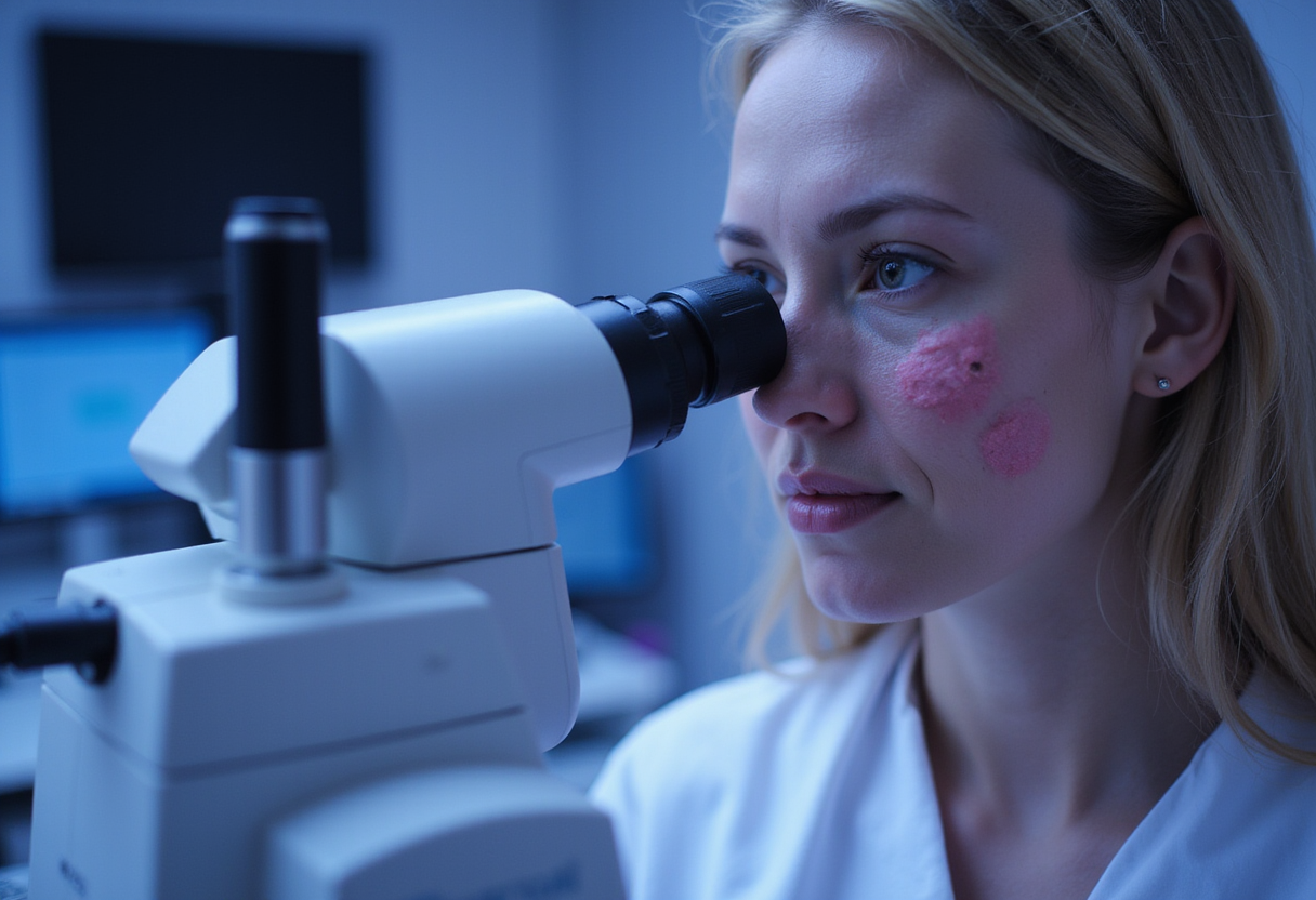
Artificial intelligence combined with Raman spectroscopy improves the detection of skin cancer, according to a report published by Technology Networks — a pairing that targets one of the most persistent gaps in dermatologic diagnosis: distinguishing malignant from benign lesions without a biopsy.
The core idea is mechanical, not cosmetic. Raman spectroscopy measures how light scatters when it strikes tissue. Because molecular components of skin — lipids, proteins, nucleic acids — shift scattered photons in characteristic ways, the technique produces a biochemical fingerprint of a lesion. Cancerous tissue has a different molecular composition than healthy skin, and those differences show up in the Raman signal.
That signal, however, is subtle. Raw spectra are noisy, overlapping and difficult for a clinician to read at the bedside. This is where the machine-learning layer enters. An AI model trained on labeled spectra learns which patterns of molecular vibration correlate with malignancy and which do not. The reported improvement in detection performance comes from this division of labor: the spectrometer supplies objective biochemical data, and the algorithm interprets it at a consistency human pattern recognition cannot match.
Why does this matter for dermatology?
Skin cancer diagnosis still hinges heavily on visual inspection — dermatoscopy where available, and excisional or punch biopsy as the decisive test. Biopsy is accurate but invasive, slow to return results, costly at scale, and a bottleneck in high-volume clinics and skin-cancer screening programs. The consequence is well known to practicing dermatologists: a large share of biopsied lesions prove benign, while some malignancies are initially missed.
A non-invasive optical tool that reduces both false positives and false negatives would change that economics. It would allow clinicians to triage lesions in real time, biopsy only where the algorithm and spectroscopic signal indicate genuine risk, and prioritize suspicious cases for faster excision. In populations with limited access to dermatologists — rural regions, underserved clinics, high-prevalence countries such as Australia — a portable spectroscopy-plus-AI device could extend diagnostic capacity beyond specialist centers.
How does the method work?
The workflow reported follows the logic of other optical diagnostic platforms:
- Signal acquisition. A Raman probe illuminates the lesion with laser light and captures the inelastically scattered photons.
- Spectral preprocessing. Software filters noise and normalizes the spectrum so lesions are comparable across patients and instruments.
- Classification. A trained AI model assigns the lesion a risk category based on the biochemical signature.
Unlike visual AI tools that train on clinical or dermoscopic images, Raman-based classification reads chemistry rather than morphology. That matters because visually ambiguous lesions — amelanotic melanomas, lesion mimics, heavily pigmented nodules — are exactly where image-based systems and clinicians struggle most.
What are the open questions?
The Technology Networks report describes improved detection but leaves several practical questions that will determine clinical adoption. Sensitivity and specificity figures in real-world cohorts, performance against expert dermatologists using dermoscopy, and validation across skin phototypes remain the benchmarks any such device must clear before regulators and payers accept it. Training data provenance is equally critical: an algorithm validated on one population's spectra may not generalize to another's without retraining.
Cost and workflow fit are not trivial either. A device must be fast enough for clinic pace, rugged enough for field screening, and cheap enough to compete with the biopsy it aims to reduce. Handheld Raman probes exist, but their diagnostic algorithms have historically required large, curated spectral databases to reach robust performance.
What comes next?
For now, the finding adds momentum to a clear trend: molecular-level, AI-interpreted diagnostics moving from the lab toward point-of-care skin-cancer triage. Dermatologists should watch for prospective multicenter validation trials — and, if those hold up, the sensitivity and specificity numbers that would justify integrating spectroscopic assessment into routine lesion evaluation.
Source: Google News: Skin cancer
Daniel Okafor
Show full bio
Staff writer covering people, launches and day-to-day practice in Clinical dermatology, inflammatory skin disease and biologics at Derm Field.